DRESDEN, Germany — The thalamus, a region of the inner brain, is made up of many divisions, each of which carries out a specific function. Ranging from interpreting and conveying impulses for sense and movement to regulating awareness and attentiveness, it is essentially the ”middle man” in the communication between the brain and our sensory organs. Neuroscientists have studied the pathways by which the thalamus helps to control the senses, however, it has been a difficult feat to determine exactly how this region works.
Now, scientists from the Dresden University of Technology, in Germany, have developed technology that allows us a sneak peek inside the interconnections of the thalamus and visual pathways. This is a critical step towards identifying damaged areas in those with vision issues.
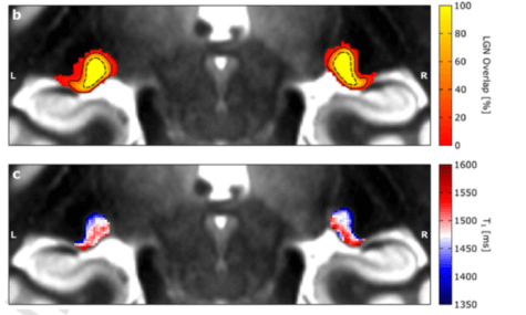
Many conditions produce symptoms of visual impairment. For example, children with dyslexia often develop visual issues. The specific area of the thalamus that controls vision works to relay signals to and from the two hemispheres of the brain and the eyes. It is divided into two principal divisions: the lateral geniculate nucleus and the striate cortex. Since both of the divisions related to visual processing are extremely small and deeply buried in the brain, scientists have had trouble evaluating them in live individuals up to this point.
Christa Müller-Axt, a Ph.D. student at Dresden, happened upon these regions while studying MRI scans of a person with dyslexia. She found patterns she believed may be similar to the sensory division of the thalamus which controls vision. According to Müller-Axt and Professor Katharina von Kriegstein, a neuroscientist at the university, the specialized MRI scans reveal data with unusually ”high spatial resolution” that was previously unseen. In other words, these areas showed up very well via MRI scan.
To be certain of the results, she used the same MRI technique in living and deceased brain tissue and examined the thalamic region of the deceased by extracting tissue samples. Müller-Axt was able to confirm that she had in fact uncovered the two primary regions which control visual input from the thalamus to the visual cortex of the brain. The findings revealed that both of these regions vary in myelin, or “white matter,” thickness making it possible to identify these regions via MRI scan.
“The finding that we can display visual sensory thalamus compartments in living humans is fantastic, as it will be a great tool for understanding visual sensory processing both in health and disease in the near future,” says CMüller-Axt, who is also the study’s first author, in a statement. “Post-mortem studies in developmental dyslexia have shown that there are alterations specifically in one of the two compartments of the visual sensory thalamus. However, there are very few of these post-mortem studies, so it is difficult to say whether all dyslexics are characterized by these kinds of visual sensory thalamus alterations.
“Also, post-mortem data cannot reveal anything about the functional impact of these alterations and their specific contribution to developmental dyslexia symptoms,” she adds. “Therefore, we expect that our novel in-vivo approach will be a great asset in facilitating research on the role of the visual sensory thalamus in developmental dyslexia.”
The new, unintrusive technique may soon give a thorough grasp of how the brain processes visual input. Moreover, additional research using this technique could lead to potential treatments for those suffering from vision impairment issues, like glaucoma and visual issues caused by dyslexia.
The study’s findings are published in the journal NeuroImage.