Researchers from Osaka University found that Ca2+ signaling simultaneously performed signal amplification and olfactory adaptation. The results demonstrated that this mysterious phenomenon was segregated inside the cilium. A novel system was used to observe changes in Ca2+ dynamics inside the thin structure of a cilium. Unveiling the mystery of Ca2+ signaling segregation further clarifies the mechanisms underlying the human sense of smell.
In a study published this month in the Journal of General Physiology, researchers revealed that the amplification and reduction of the sensory signal are both regulated by Ca2+, and the processes are clearly segregated within a tiny structure of a sensory cell.
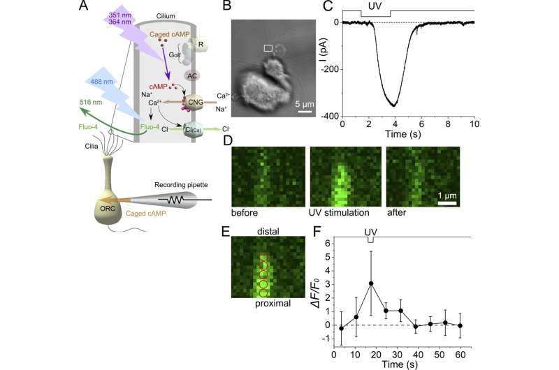
The initial event that induces the sense of smell is the binding of odorant molecules to the hair-like structures, cilia, attached to the olfactory receptor neurons in the nose. This binding triggers the flow of ions causing an electrical excitation in the cilia, controlled by ion channels that promote the influx of extracellular positive ions into the semifluid compartment of the cilia.
As a result, the Ca2+ signal simultaneously performs the opposing effects of excitation and adaptation to regulate the activity of ion channels on the cilia. A series of events leading to the initiation of a biochemical cascade promotes the increase of Ca2+ inside the cilia: the current increases, impulses are generated, and information is sent to the brain to identify the scent. In the adaptation process, negative feedback signaling occurs to prevent saturation of ion channel activities and to adjust sensitivity to stimulus intensities.

“How the same ion controls both the increase and decrease in local current has been a mystery,” explains lead author Hiroko Takeuchi. “In this research, we aimed to determine whether the process of signal amplification and reduction is indeed segregated only inside the cilia.” The phenomenon remains a mystery because of the technical difficulty in observing the change in Ca2+ dynamics inside the thin structure of the cilia.
UV excitation of the local cilium immediately generated the inward current and Ca2+ signal; however, after the termination of the stimulus, the current decreased together with the reduction of Ca2+ signal. Surprisingly, however, the adaptation persisted for longer periods even after the Ca2+ signal disappeared. It seems likely that Ca2+ is kept bound exclusively to proteins that regulate the adaptation. “Our results were promising; these opposing effects of Ca2+ occurred separately by molecular kinetics in a tiny space of the native cilium,” says senior author Takashi Kurahashi.
The successful capture of ion channel activity and Ca2+ dynamics within the cilia is a key step toward understanding the human sense of smell. By elucidating the intricacies of Ca2+ signaling, the human sense of smell could be enhanced through the development of olfactory sensors or adjustments to the chemical environment of the nose.
The article, “Segregation of Ca2+ signaling in olfactory signal transduction,” was published in the Journal of General Physiology.
More information: Hiroko Takeuchi et al, Segregation of Ca2+ signaling in olfactory signal transduction, Journal of General Physiology (2023). DOI: 10.1085/jgp.202213165
Journal information: Journal of General Physiology
Provided by Osaka University
Source: Researchers identify cellular mechanism that improves olfactory function